

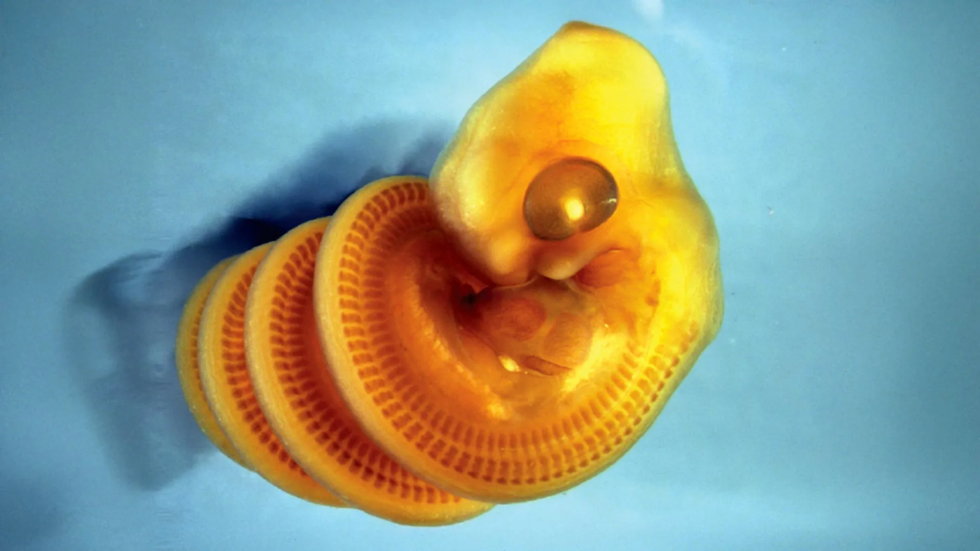
An international research team led by scientists at the Canadian Museum of Nature has identified the mechanism behind why snake embryos develop in tight spiral formations before hatching. The findings, published in Current Biology, reveal that the coiling behavior enables snakes to accommodate their distinctively elongated bodies relative to other vertebrates. The spiral pattern forms as the embryo’s body grows rapidly while the gut remains comparatively static, creating a physical constraint that causes the lengthening body to buckle and twist into a right-handed coil, similar to how a strap twists when adjusted unevenly.
The research project originated during a period of laboratory restrictions in 2020, when Dr. Tetsuto Miyashita sought a research question that could be pursued remotely. The team, including lead author Alexandra Weber, examined photographs of more than 900 snake embryos representing 39 different snake and limbless squamate species collected from published research and museum databases. The analysis revealed a consistent pattern: during early developmental stages, all embryos displayed right-handed coiling when viewed from head to tail. Since the embryos lacked developed muscles at these stages, the researchers concluded that physical developmental forces rather than muscular movement were responsible for the twisting pattern.
CT imaging conducted by collaborator Dr. Raul Diaz provided the crucial anatomical evidence. The scans revealed an unusual structure within developing snake embryos: a pillar of intestinal tissue stretching through the spiral, surrounded by blood vessel tendrils from the yolk sac. This discovery explained the mechanical mechanism: the mismatch between the rapid lengthening of the body and the slower growth of the gut creates a constraint that forces the body into a spiral configuration. The positioning of the yolk sac on the left side of the embryo dictates that coiling always begins in a right-handed direction.
As snake embryos continue developing, their relationship to this spiral configuration changes. The yolk sac shrinks, providing more spatial freedom, and the embryo’s muscles mature sufficiently to enable active movement. Consequently, some embryos remain right-handed while others reorient to left-handed coiling by the time they approach hatching. The research suggests that this directional shift reflects the transition from passive mechanical constraint to active muscular control during fetal development.
The team proposes that this mechanical model of spiral formation could provide insights into other spiral structures observed throughout nature, including intestinal looping in mammals and shell formation in mollusks. The discovery exemplifies how fundamental biological questions can be addressed through systematic observational analysis rather than solely through molecular genetic approaches.
Article Attribution | Read More at Article Source
Article summary produced by Claude AI