

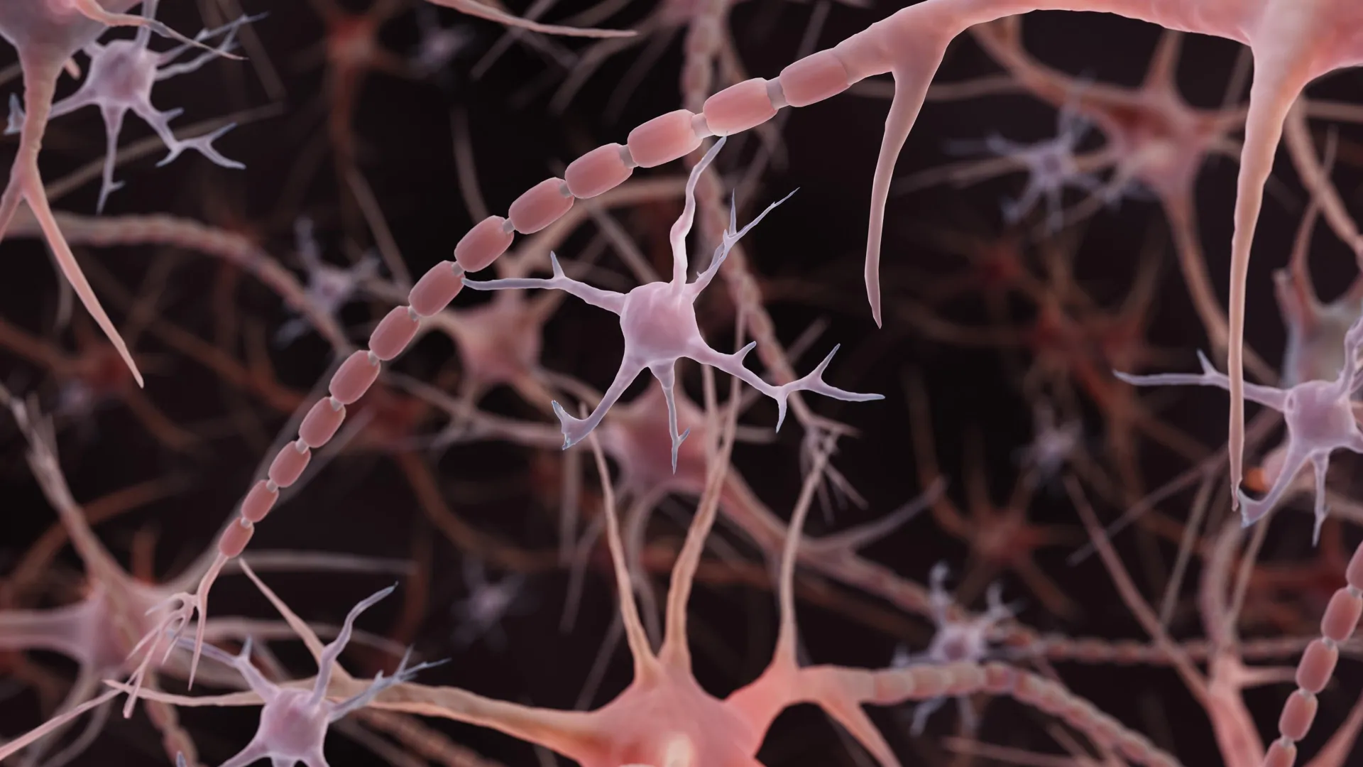
Scientists at Johns Hopkins Medicine have identified a previously underappreciated structural feature of axons, the signal-carrying extensions of brain cells, according to research published in Nature Neuroscience on December 2, 2024. Rather than the smooth, uniform tubes depicted in standard textbook illustrations, axons appear to contain repeating small bulges resembling strings of pearls. These structures, termed non-synaptic varicosities, are distinct from the larger bulges at communication sites between neurons and may represent a normal feature of axon architecture rather than a sign of cellular damage.
The investigation began when researcher Shigeki Watanabe noticed similar pearled structures in worm neurons and collaborated with colleagues to explore the phenomenon more broadly. Graduate student Jacqueline Griswold and the team used high-pressure freezing electron microscopy to examine neurons from laboratory cultures and mouse tissue samples. This preservation method proved critical, as it maintained the delicate cellular structures without the distortions that can result from standard chemical fixing and dehydration techniques. The team observed consistent pearling patterns across tens of thousands of tissue images and confirmed the structures were present in living neurons as well.
To understand the mechanism behind these pearled axons, the researchers developed mathematical models of the membrane surrounding the axons and collaborated with theoretical biophysicist Padmini Rangamani. The models suggested that physical forces and membrane properties alone could explain the pearl-like appearance without requiring internal structural templates. Experiments manipulating sugar concentration and cholesterol content altered the size and shape of the pearls, supporting the role of membrane mechanics. Related research has since documented these pearled structures in human brain tissue, extending the findings beyond mice.
The researchers further discovered that neural activity itself can reshape these structures. Following high-frequency electrical stimulation, the pearl-like regions enlarged by an average of 8% in length and 17% in width, with changes persisting for at least 30 minutes. These structural modifications appeared to influence the speed of electrical signal transmission, with larger pearls affecting ion movement through the axon. The findings suggest that axons function not as static cables but as dynamic structures capable of responding to neural activity and modulating signal transmission properties.
Article Attribution | Read More at Article Source
Article summary produced by Claude AI