

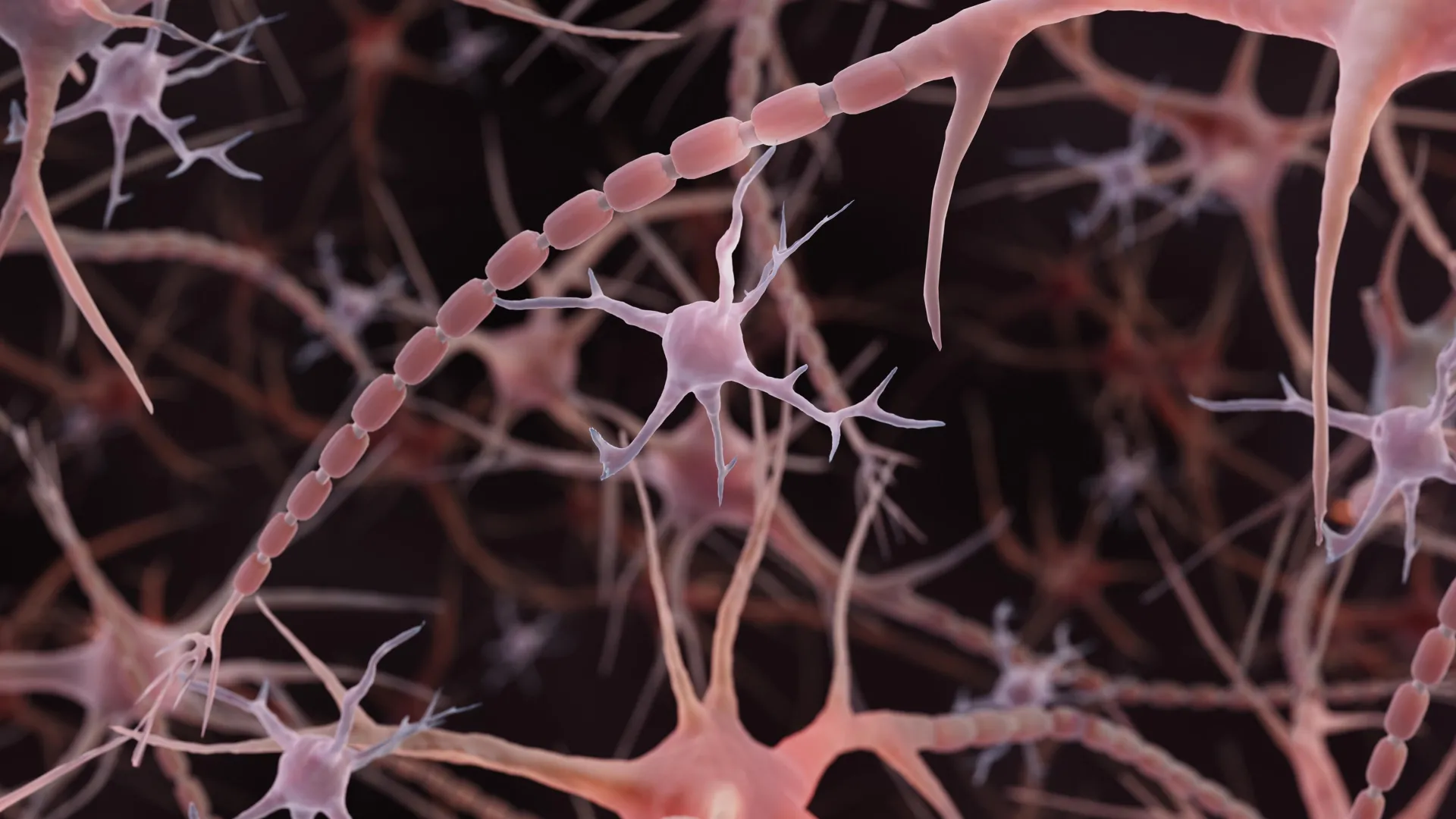
A study by Johns Hopkins Medicine published in December 2024 reveals that axons, the long extensions through which neurons transmit electrical signals, may possess a previously overlooked structural feature. Rather than the smooth, uniform tubes depicted in conventional textbook illustrations, some axons display repeating bulges resembling strings of tiny pearls. These structures, termed “non-synaptic varicosities,” appear to be part of normal axon architecture rather than signs of cellular damage, as scientists had previously assumed when observing pronounced beading in diseased neurons.
The research began with observations of repeating pearled patterns along axons in worms, prompting further investigation into whether this architecture occurs in mammalian neurons. Using high-pressure freezing electron microscopy—a technique that preserves delicate cellular structures better than conventional chemical fixation—the researchers examined thousands of images of mouse neurons in various states. The pearled appearance remained consistent across laboratory-grown neurons, adult neurons, and embryonic neurons, and the pattern persisted even in living neurons observed with high-resolution imaging, confirming it was not an artifact of the freezing process.
To understand the mechanism behind this structure, the team developed mathematical models of axon membranes and conducted experiments manipulating membrane properties. By adjusting sugar concentration and cholesterol levels, they demonstrated that physical forces acting on the membrane could produce the pearled pattern without requiring a rigid internal framework. These findings suggested that the axon’s shape is not fixed but responsive to its chemical and physical environment.
The research further demonstrated that electrical activity can reshape these structures, with high-frequency stimulation causing the pearl-like regions to enlarge by approximately 8% in length and 17% in width, with effects persisting for at least 30 minutes. This activity-dependent remodeling correlated with changes in electrical signal transmission speed. Subsequent studies confirmed the presence of similar pearled axons in human brain tissue, extending the observations beyond laboratory mouse models and suggesting this structural feature may be fundamental to neural organization across mammalian species.
Article Attribution | Read More at Article Source
Article summary produced by Claude AI