

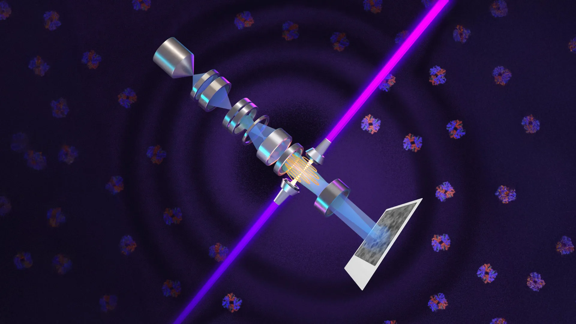
Researchers at the University of California, Berkeley have created a new imaging device called a laser phase plate designed to enhance cryoelectron microscopy (cryo-EM) and cryoelectron tomography (cryo-ET). The innovation applies a century-old microscopy principle to modern electron imaging technology, potentially addressing a major limitation in studying biological structures.
The laser phase plate uses what researchers describe as the world’s most intense focused continuous-wave laser to interact directly with an electron beam, altering its phase to increase image contrast. This enhancement could enable scientists to image proteins approximately one-third the size of those currently observable, while also improving visibility of cellular structures such as the nucleus and mitochondria. The technology is particularly significant for cryo-ET, which constructs three-dimensional views of proteins and molecules from multiple angles and can reveal how biological structures interact within their natural cellular environment.
The development builds upon work initiated in 2010 when physicist Holger Müller and colleague Robert Glaeser proposed using intense lasers to shift electron beam phases without compromising beam intensity. Müller, a UC Berkeley professor of physics, led the current effort. The breakthrough was made possible through financial support from Biohub, a research organization in Redwood City, California. Two research instruments have been developed: one at UC Berkeley, named Theia after the ancient Greek Titaness associated with light, and another at Biohub’s imaging facility that incorporates two lasers arranged at right angles to improve stability and reduce optical distortion.
The laser phase plate technology addresses challenges that have long constrained biological imaging. Because cellular material is largely transparent and small structures scatter minimal light, scientists have historically struggled to achieve sufficient contrast for detailed observation. The new approach applies principles from phase-contrast microscopy, a technique developed in 1930 and recognized with a Nobel Prize in 1953, to electron microscopy. Both research teams are collaborating with Thermo Fisher Scientific, the leading manufacturer of cryo-EM instruments. The Berkeley team’s findings were published in the journal Science, while Biohub’s two-laser design details appear in a recently posted preprint.
Article Attribution | Read More at Article Source
Article summary produced by Claude AI