

Researchers at Stanford University have created an advanced imaging system called Interferometric Image Scanning Microscopy (iISM) that visualizes the interior of living cells with unprecedented clarity and detail. The technology merges two distinct microscopy approaches to achieve a resolution of approximately 120 nanometers while maintaining the ability to observe cells without the fluorescent dyes traditionally required to make cellular structures visible. The innovation was described in the Nature portfolio journal Light: Science and Applications and represents a significant advancement in biological imaging capabilities.
A primary advantage of the new microscope lies in its ability to image cells without fluorescent markers. Conventional microscopy techniques typically rely on fluorescent substances to highlight specific cellular structures, a method that carries notable limitations. These approaches generally allow researchers to label only a small number of structures simultaneously, making it difficult to observe how different cellular components interact. Additionally, fluorescent signals diminish over time through a process called photobleaching, and introducing labels can be challenging and potentially alter the behavior of the structures being studied. The Stanford system circumvents these challenges by operating without such markers, enabling extended observation of numerous cellular components in a more natural state. The technology also requires substantially less illumination power than comparable label-free methods, reducing the risk of photodamage that can harm living cells or interfere with their normal functions.
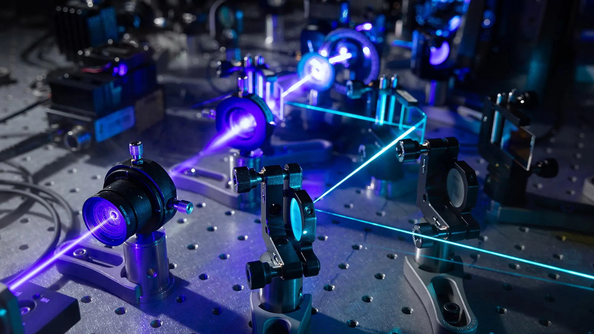
The breakthrough results from combining interferometric scattering microscopy with detector array technology adapted from advanced confocal microscopes. In interferometric scattering microscopy, a laser illuminates a cell, causing internal structures to scatter light; a second laser beam amplifies this weak signal through interference. The team enhanced this approach by incorporating an array detector capable of collecting tens to hundreds of views simultaneously, rather than the single perspective captured by traditional systems. A computational method then merges these multiple measurements to produce images with enhanced sharpness, contrast, and depth information. The result is an instrument capable of resolving extremely small cellular structures while maintaining imaging speed and minimizing cell damage.
Researchers emphasize that iISM is intended to complement rather than replace fluorescence microscopy. The technology could advance cancer research, drug development, infectious disease studies, and investigations of plant-microorganism interactions. Scientists can now investigate cellular responses to drugs and pathogens, potentially uncovering biological processes previously difficult to study.
Article Attribution | Read More at Article Source
Article summary produced by Claude AI