

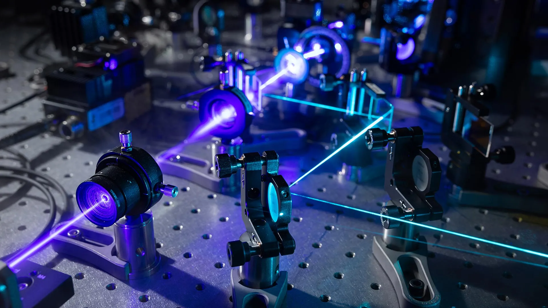
Researchers at Stanford University have unveiled an advanced microscope system that provides detailed imaging of living cells without requiring fluorescent markers. The instrument, called Interferometric Image Scanning Microscopy (iISM), achieves a resolution of approximately 120 nanometers by combining two microscopy techniques. The development was published in the Nature portfolio journal Light: Science and Applications and could advance cancer research, drug development, and infectious disease studies.
A primary advantage of the new system is its ability to visualize cellular structures without fluorescent labels, which traditional microscopy techniques typically require. Conventional fluorescence methods can only label a limited number of structures simultaneously, experience signal degradation through photobleaching, and may alter the behavior of observed components. The Stanford system eliminates these constraints by allowing researchers to follow numerous cellular components over extended periods while maintaining a more natural biological environment. Additionally, iISM requires substantially less illumination power than comparable label-free methods, reducing photodamage that can harm cells or interfere with their normal functions.
The breakthrough results from combining interferometric scattering microscopy with detector array technology adapted from advanced confocal microscopes. Interferometric scattering microscopy exploits how light scatters when encountering tiny particles within cells. A laser illuminates the cell, and scattered light is amplified through interference using a second laser beam. Researchers enhanced this approach by incorporating an array detector that collects tens to hundreds of views from slightly different perspectives, similar to how human stereo vision provides depth perception.
Computational methods then combine these multiple measurements into sharper, higher-contrast images with improved depth information and precision. The detector array collects more light than traditional single-detector systems, enabling the 120-nanometer resolution without sacrificing imaging speed or requiring intense illumination. This allows scientists to observe living cells for extended periods while minimizing disturbance or damage. Researchers note that iISM is designed to complement rather than replace fluorescence microscopy, with the combined approach potentially addressing previously difficult research questions.
Article Attribution | Read More at Article Source
Article summary produced by Claude AI