

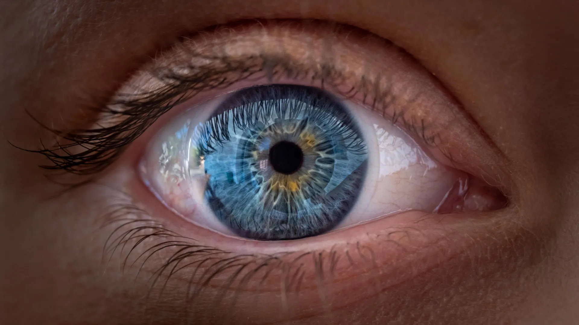
Scientists at Johns Hopkins University have identified previously unknown mechanisms underlying the development of human vision, specifically how specialized light-detecting cells arrange themselves in the foveola, the region of the retina responsible for sharp central sight. The research, published in Proceedings of the National Academy of Sciences, demonstrates the coordinated roles of a vitamin A derivative and thyroid hormones in shaping the eye’s structure before birth. The findings contradict a longstanding explanation in vision science and may inform future treatments for conditions such as macular degeneration and glaucoma.
To conduct their investigation, researchers led by associate professor Robert J. Johnston Jr. employed laboratory-grown miniature versions of human retinal tissue called organoids. Created from fetal cells, these structures reproduce key characteristics of developing human tissue and allowed scientists to observe changes over several months that would be difficult to track during pregnancy. The team focused particularly on the foveola, a tiny central retinal region that comprises roughly 50% of human visual perception despite occupying only a small portion of the retina. The foveola’s exceptional role in detailed central vision—enabling reading, facial recognition, and perception of fine details—depends on a specific arrangement of cone cells that detect different wavelengths of light.
The researchers identified two chemical processes that shape cone cell distribution in the foveola. Between weeks 10 and 12 of fetal development, blue cones initially appear in this region. By week 14, evidence suggests these cells have transformed into red and green cones. Retinoic acid, a molecule derived from vitamin A, limits blue cone formation in the central retina, while thyroid hormones appear to convert remaining blue cones into red or green variants. This coordinated process creates the precise arrangement necessary for optimal vision, as blue cones in the foveola would compromise visual acuity.
These findings challenge a model that has dominated vision research for approximately 30 years. The prevailing theory held that blue cones present in the foveola during development simply migrated away as the eye matured, with cone cell types remaining fixed once established. The new evidence suggests instead that at least some blue cones may change their fundamental identity rather than relocate. Johnston emphasized that the research describes vitamin A’s role in prenatal eye development and does not suggest that vitamin A supplements can enhance vision in adults. The research team hopes the findings may eventually support development of laboratory-grown retinal tissues for transplantation to restore vision in patients experiencing sight loss.
Article Attribution | Read More at Article Source
Article summary produced by Claude AI